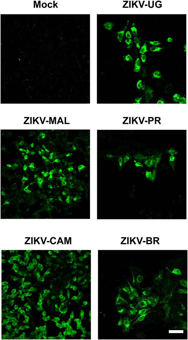
Figure 4.
Infection of differentiated SH-SY5Y cells with ZIKV strains representing African and multiple Asian lineages. Viral infection is monitored by immunofluorescent staining of differentiated SH-SY5Y cells at 48 hours post-infection using the anti-Env monoclonal antibody (green). Cells are treated with 2 days of retinoic acid prior to ZIKV infection. The images shown are representative images taken from 3 independent experiments, with observation of a minimum of 10 fields under both low (10X) and high (40X and 63X) magnification per experiment. The scale bar represents 50 µm.

