Fig. 1.
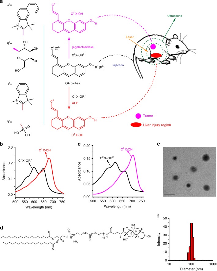
Schematic illustration for the probes’ response in vivo and related properties. a Schematic illustration for two xanthene-based molecules (C1X-OR1 and C2X-OR2) as the activatable OA probes for respectively imaging liver injury and metastatic tumor. b and c Absorption spectra for the probes (5 μM) and their corresponding activated forms (C1X-OH and C2X-OH, 3 μM). d Structure of a hepatocyte-targeting phospholipid for liver targeting. e and f Transmission electron microscopic image and particle diameter distribution for a liposomal C1X-OR1 sample. Scale bar: 200 nm

