Figure 1.

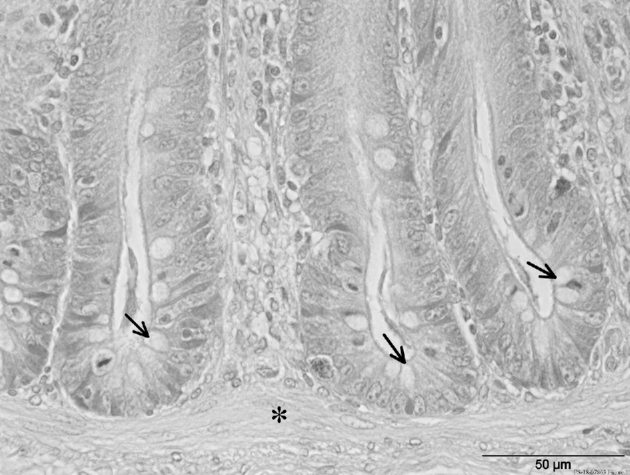
H&E staining from cecal crypts (apex region) of broiler chickens with SP diet. Note especially the goblet cells (arrows) within crypts and the lamina muscularis mucosae (star) underneath. Scale bars = 50 μm.

H&E staining from cecal crypts (apex region) of broiler chickens with SP diet. Note especially the goblet cells (arrows) within crypts and the lamina muscularis mucosae (star) underneath. Scale bars = 50 μm.