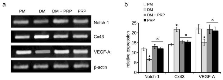
Figure 3.
RT-PCR analysis of Notch-1, Connexin 43 (Cx43), and VEGF-A expression. NIH/3T3 fibroblastic cells were cultured in differentiation medium (DM, low serum medium plus 2 ng/mL TGF-β1) in the absence or presence of 1:50 diluted PRP (DM + PRP) or in the presence of 1:50 serum-free medium diluted PRP (PRP). Cells cultured in proliferation medium (PM) served as controls, undifferentiated cells. (a) Representative agarose gels. (b) Histogram showing the densitometric analyses of the bands normalized to β-actin. Data shown are mean ± S.E.M. and represent the results of at least three independent experiments performed in triplicate. Significance of difference: * p < 0.05 vs. PM; ° p < 0.05 vs. DM.

