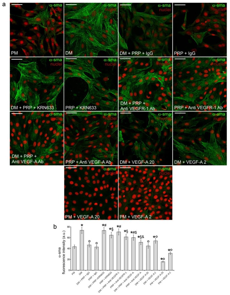
Figure 5.
Effect of VEGFR-1 inhibition, VEGF-A neutralization, and stimulation with soluble VEGF-A on α-sma expression: confocal immunofluorescence analysis. NIH/3T3 fibroblastic cells were cultured in differentiation medium (DM, low serum medium plus 2 ng/mL TGF-β1) in the absence or presence of 1:50 diluted PRP + irrelevant IgG (DM + PRP + IgG) or in the presence of 1:50 serum-free medium diluted PRP + IgG. To evaluate the involvement of VEGF-A/VEGFR-1 mediated signaling in PRP-induced fibroblast response, the cells were treated with the selective pharmacological VEGFR inhibitor, KRN633 (DM + PRP + KRN633; PRP + KRN633) or with anti-VEGFR-1 neutralizing antibodies (8 µg/mL; DM + PRP+ Anti VEGFR-1 Ab; PRP + Anti VEGFR-1 Ab) or anti-VEGF-A neutralizing antibodies (10 µg/mL ; DM + PRP+ Anti VEGF-A Ab; PRP + Anti VEGF-A Ab). In parallel experiments the cells were cultured in DM or PM in the presence of two different concentrations of soluble VEGF-A (20 ng/mL, DM + VEGF-A 20, PM + VEGF-A 20; 2 ng/mL, DM + VEGF-A 2, PM + VEGF-A 2). Cells cultured in proliferation medium (PM) served as control undifferentiated cells. (a) Representative confocal fluorescence images of the cells immunostained with antibodies against α-sma and counterstained with propidium iodide to reveal nuclei. Scale bar: 50 µm. (b) Histogram showing the densitometric analysis of the intensity of α-sma fluorescence signal performed on digitized images. Data shown are mean ± S.E.M. and represent the results of at least three independent experiments performed in triplicate. Significance of difference: * p < 0.05 vs. PM; ° p < 0.05 vs. DM; # p < 0.05 vs. DM + PRP + IgG; § p < 0.05 vs. PRP + IgG; $ p < 0.05 vs. DM + PRP + Anti VEGFR-1 Ab; & p < 0.05 vs. PRP + Anti VEGFR-1 Ab.

