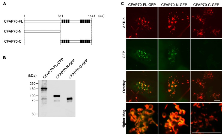
Figure 3.
Changes in the ciliary localization of CFAP70 fragments in primary culture of mouse ependyma. (A) Schematic diagrams of mouse CFAP70 protein and its deletion constructs made in this study. The 1141-amino-acid protein has clusters of tetratricopeptide repeat (TPR) domains (indicated by filled boxes) at the C-terminal half. (B) Western blot analysis for validation of the expression of recombinant CFAP70 proteins fused with a GFP tag at the C-terminus. 293T cells were transfected with the lentiviral expression plasmids and the whole cell lysates were analyzed. The calculated molecular weights are as follows: CFAP70-FL-GFP, 155 kDa; CFAP70-N-GFP, 95 kDa; CFAP70-C-GFP, 87 kDa. To the left of the gel, the positions and sizes (kDa) of molecular weight standards are indicated. (C) Confocal microscopy of cultured ependyma transduced with lentiviral particles carrying the above constructs. The cells were immunostained for AcTub (red) and GFP (green). Bars, 10 µm.

