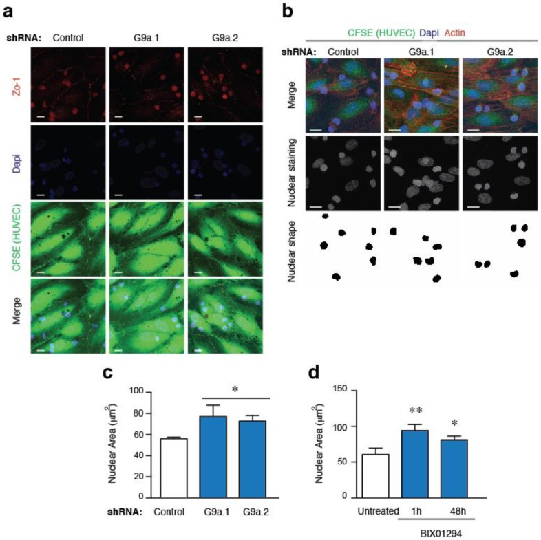
Figure 2.
G9a depletion increases the nuclear area of ALL cells. (a) HUVEC cells were grown to confluency, labelled with CFSE and stimulated with TNFα for 16 h. Control or G9a depleted Jurkat cells were plated on TNFα-activated HUVEC cells. Cells were fixed, permeabilized and analyzed to visualized their nuclei (DAPI, blue), F-actin (Phalloidin, cyan), and endothelial junctions (Zo-1, red); (b) Control and G9a depleted Jurkat cells were cultured on CFSE labelled HUVEC activated with TNFα, fixed and stained for DAPI (blue) and F-actin (red). Nuclear shapes were determined; (c) Graph shows the nuclear areas quantified from (b); Mean n = 3 replicates ± SD. Bar = 10 μm. * p < 0.05; (d) Graph shows the nuclear areas from untreated or BIX10924 treated Jurkat at cells cultured on TNFα-activated HUVEC. Mean n = 3 replicates ± SD. * p < 0.05; ** p < 0.01.

