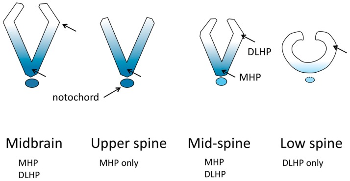
Figure 2.
Conceptual views of the shape of neuroepithelium in transverse sections, at various anterior–posterior locations, of a mouse neural tube before closure, demonstrating differences in bending mechanisms. Shading represents the concentration of Shh signaling. DLHP, dorsolateral hinge point. MHP, medial hinge point. Modified from Figure 3 in [13].

