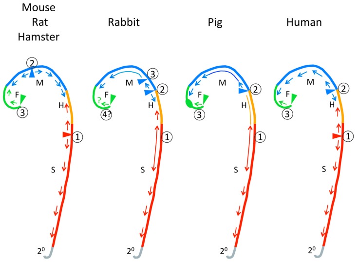
Figure 3.
An interpretation of the patterns of neural tube closure in various mammalian species based on published images and studies. Colors denote future anterior–posterior fates of the neural tube. F, forebrain; M, midbrain; H, hindbrain; S, spine. Short arrows indicate direction of zipping. Long stems on arrows and lines lacking arrowheads denote areas that appose and then fuse simultaneously. Triangles and circled numbers indicate closure initiation sites. The forebrain oblong denotes a region that appears to close from all sides, rather than apposition or zipping. 2°, region of secondary neurulation.

