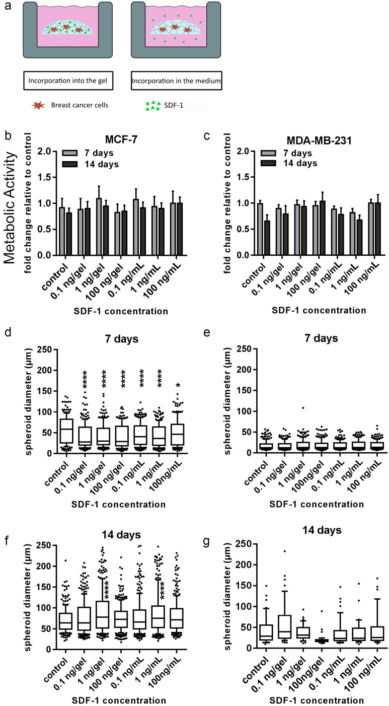
Figure 7.
Cell viability and tumor spheroid diameter of MCF-7 and MDA-MB-231 cells when exposed to stromal cell-derived factor 1 (SDF-1). (a) SDF-1 was incorporated into either the gel or in the media for 14 d of culture. PrestoBlue assays and microscopic analyses were performed. (b,c) Viability data are presented as fold change relative to untreated control (±SEM). (d–g) Box plot data represent median values, percentiles (10%–90%), and outliers of spheroid diameters of MCF-7 cells at 7 d (d) and 14 d (f) and MDA-MB-231 cells at 7 d (e) and 14 d (g). Experiments were performed three times in triplicate (n = 3). Asterisks (*) denote statistical significance: * (p < 0.05), ** (p < 0.01), *** (p < 0.001) or **** (p < 0.0001) from the control samples.

