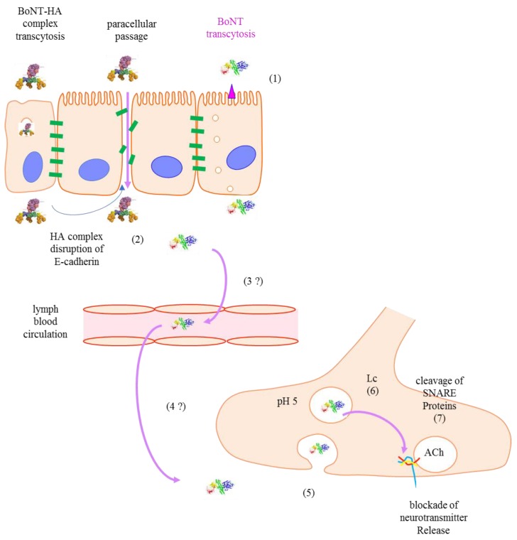
Figure 2.
Route and mechanism of intoxication by BoNT. (1) BoNTs and BoNT-hemagglutinin (HA) are transcytosed through epithelial and microfold (M) cells respectively in human intestinal epithelia; (2) In the extracellular region, the trimeric HA complexes bind to E-cadherin, leading to intestinal barrier disruption and paracellular passage; (3 and 4) BoNTs enter into the circulatory system and reach nerve terminals by an unknown way; (5) BoNTs bind to polysialoganglioside (PSG) and synaptic vesicle glycoprotein 2 (SV2); and enter neurons via clathrin-dependent endocytosis; (6) Hn domains form translocation channels for Lc to escape the endosomal acidic environment toward the neutral cytosol where the Lc is activated as a Zn2+-dependent protease; (7) The Lc protease cleaves SNAP-25 with extremely high specificity to block the synaptic vesicle traffic and neurotransmitter release.

