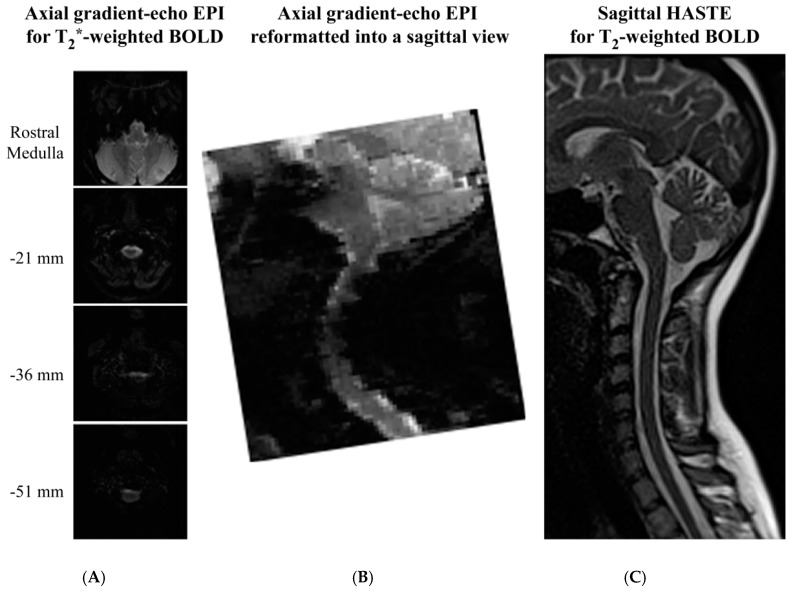
Figure 4.
Comparison of image quality obtained with gradient-echo EPI and spin-echo HASTE sequences for spinal fMRI. Gradient-echo EPI images were acquired in contiguous axial slices (A) and were reformatted into sagittal views (B) for comparison with spin-echo (HASTE) images acquired in sagittal planes (C). Selected axial slices are shown for comparison, and the slice positions are indicated relative to the caudal medulla (top slice). Images were acquired at 3 tesla with a Siemens MAGNETOM Trio at Queen’s University, and used as examples for this review.

