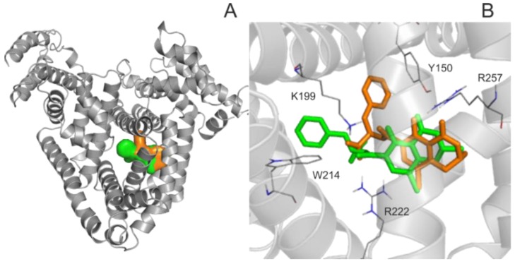
Figure 2.
Binding site and position of 2′R-OTA verses OTA in Human serum albumin (HSA). (A) HSA is represented with grey cartoon, and ligand conformation bound to Sudlow’s Site I are orange (2′R-OTA−) and green (2′S-OTA−). (B) The binding differences observed between 2′R-OTA− (orange) and 2′S-OTA− (green). The ligands are represented with colored thin sticks, while the protein binding site with grey cartoon. Hydrogen atoms of the docked ligand conformations are not presented.

