Fig. 1.
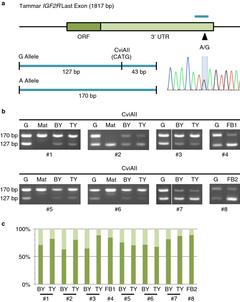
Imprinting analysis of tammar IGF2R. a Single nucleotide polymorphism in tammar IGF2R. The dark and light green parts in the box represent ORF and 3′ UTR of the last exon of tammar IGF2R, respectively. The black arrowhead and light blue line show the position of polymorphic site and the region of PCR amplification that were used for allelic expression analysis by RFLP analysis, respectively. The 170 bp PCR product contains a recognition sequence of CviAII restriction enzyme for G allele but not for A allele, enabling RFLP analysis. The sequencing data of a heterozygous sample show double peak of A and G at the polymorphic site. b Allelic expression pattern of tammar IGF2R. The intensity of 170 and 127 bp bands shows expression level from A allele and G allele, respectively. G genomic DNA, Mat maternal genotype, BY yolk sac placenta (bilaminar region), TY yolk sac placenta (trilaminar region), FB fibroblast cell line, #; individual number. c Allelic expression ratio of tammar IGF2R. The dark and light green bars represent expression ratio of active and inactive alleles, respectively. Each ratio was calculated under the normalisation using genomic DNA data as 50%

