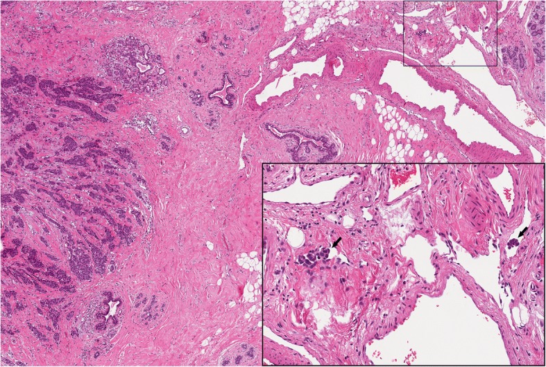
Fig. 2.

Morphological features of lymphovascular invasion in a patient with breast cancer related lymphoedema after surgery. Representative micrographs of a moderately differentiated invasive carcinoma of no special type showing peritumoral cluster of neoplastic cells inside the lumen of small vessels, as highlighted by the arrows in the inset on the bottom right. One of the two metastatic clusters determined partial lumen obliteration. H&E, original magnification × 100, inset × 400
