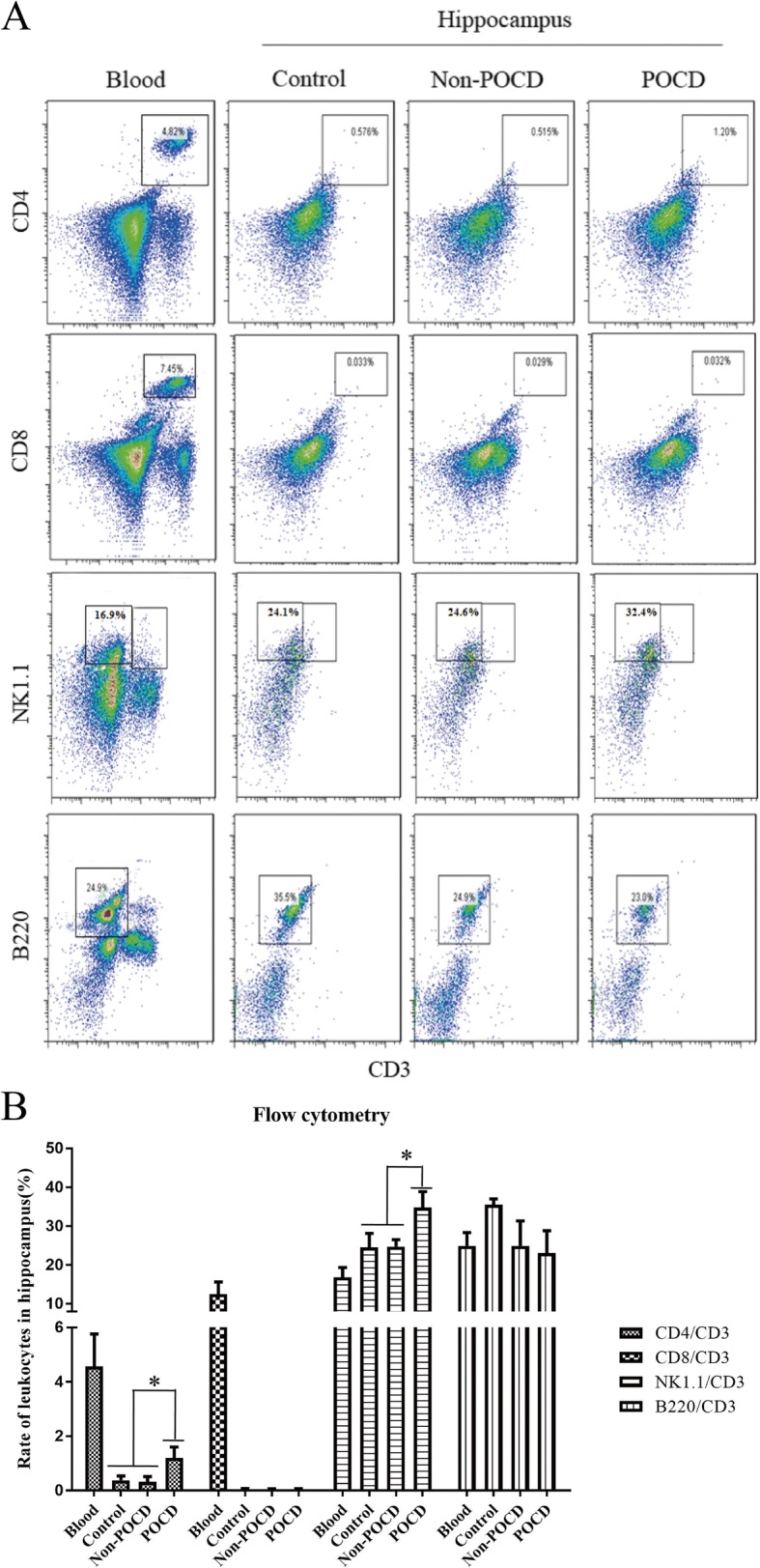
Fig. 3.

Peripheral leukocytes in hippocampus of POCD calculated using flow cytometry. Flow cytometry sorting (a) and statistic analysis (b) of leukocytes in hippocampus. “*” represents statistical significance

Peripheral leukocytes in hippocampus of POCD calculated using flow cytometry. Flow cytometry sorting (a) and statistic analysis (b) of leukocytes in hippocampus. “*” represents statistical significance