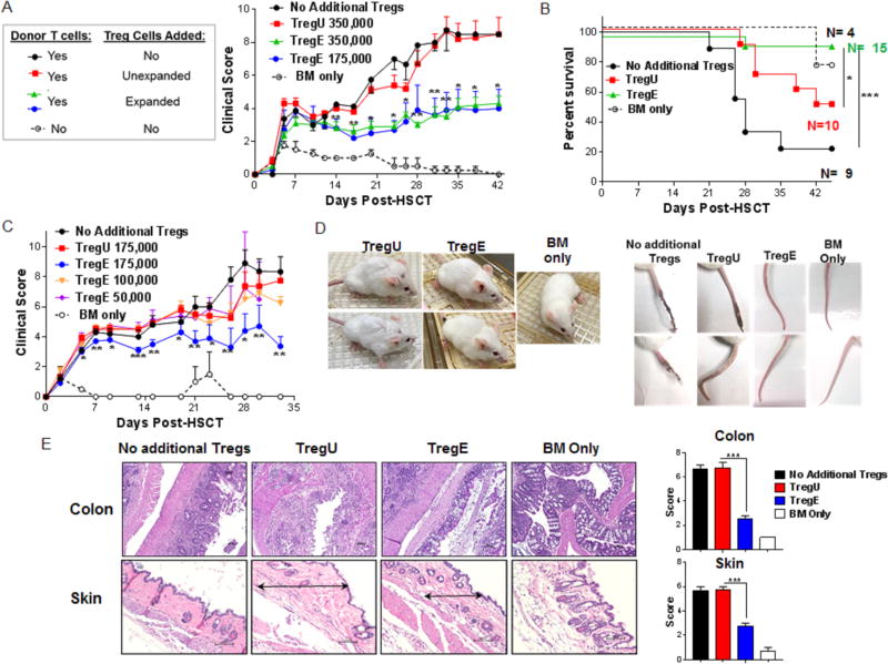
Figure 2. Low numbers of sorted TL1A-Ig+IL-2 expanded Tregs ameliorate GVHD and promote survival after MHC-mismatched aHSCT.
Tregs were expanded with TL1A-Ig+IL-2, mice were sacrificed at day 7 and splenic CD4+FoxP3+ Tregs were isolated by FACS. A complete MHC-mismatched aHSCT model (B6→ BALB/c) was utilized and varying numbers of sorted CD4+FoxP3+ unexpanded and expanded Tregs together with B6-wt 1×10⁶ splenic T cells and 5.5×10⁶ TCD B6-CD45.1 BM cells were transplanted. (A) Clinical GVHD scores (0=no disease and 10=severe) and (B) survival curves after receiving BM cells + T cells + unexpanded 350,000 (TregU), expanded 175,000/350,000 (TregE) Tregs or no addition of Tregs. BM only group was used as a negative GVHD control (n=8 mice/group, n=3 mice/BM Only group). Only recipients of “two-pathway” expanded Tregs demonstrated ameliorated GVHD. (C) Clinical GVHD scores of mice receiving unexpanded (TregU)175,000 or expanded (TregE) ≤ 175,000 Tregs corroborated the ability of 175,000 Treg E (however not lower) but not TregU to inhibit GVHD (n=6 mice/group). (D) Representative photographs of recipient mice (4 weeks post-aHSCT) and tails (9 weeks post-aHSCT) of TregU and Treg E 175,000 recipient mice illustrating healthier outcome of TregE transplanted mice. (E) Representative H&E-stained sections of colon and skin from the indicated groups 5 weeks post-aHSCT are shown. Colons from TregE 175,000 recipients showed a mild and patchy inflammation and no distortion of the villi compared with TregU 175,000 (and the no additional Treg) recipient colons which exhibited mucosal thickening, severe and extensive inflammation and edema. In skin of TregE recipients, note minimal dermal thickening (black arrow) with mild cell infiltrate compared with TregU recipients which exhibited a thickened dermis (black arrow) with extensive collagen deposition and moderate infiltration of mononuclear cells. Magnification 100× for colon and 200× for skin. Pathology scores for these tissues are shown on the right. (A-C) Data are expressed as means ± SEM and were analyzed by (A,C) a two-tailed unpaired t test or (B) log-rank test. *p<0.05; **p<0.01; ***p<0.001.

