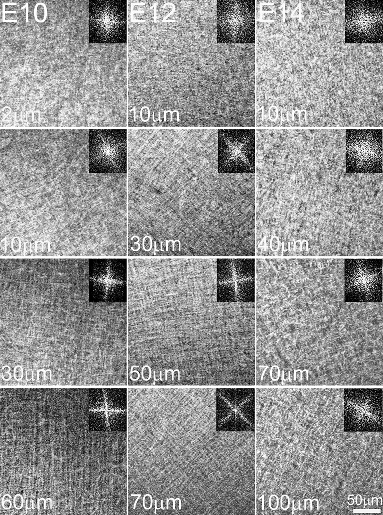
Figure 8: Keratocytes with an initially random orientation acquire an orthogonal one by E9 and display angular displacement in the anterior stroma.
Keratocyte organization in the embryonic chick corneal stroma was investigated by confocal microscopy. (A) Maximum intensity projections of en block datasets of the embryonic chick corneal stroma showing staining of actin (Phalloidin; green) and nuclei (Propidium Iodide; red). At E10, keratocytes fully invade the primary stroma. Arrowheads denote the epithelial basal lamina. Scale bar: 50μm (B) At E8, two dimensional, en face images show that most of the keratocytes have a random orientation in the anterior-mid stroma, but favour a parallel orientation in the posterior stroma aligned to the choroid fissure axis. At E9, keratocytes in the anterior stroma displayed a clockwise rotation (59.6 ± 7.1º) and orthogonal organization. Representative images from four samples per embryonic day and the respective FFT analyses at the interface are shown (insets). Scale bar: 50μm

