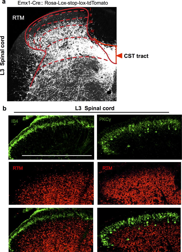
Extended Data Figure 2. CST axon termination in the lumbar spinal cord.

a-b, Representative transverse spinal section (L3) from an Emx1-tdTomato (red) reporter line (a). Sections were co-stained with IB4 (green), a lamina IIi marker and anti- PKCγ, a laminae IIi/III marker in the spinal dorsal horn (b). Scale bar: 500 μm. For a, b, 3 and 4 animals showed similar results, respectively.
