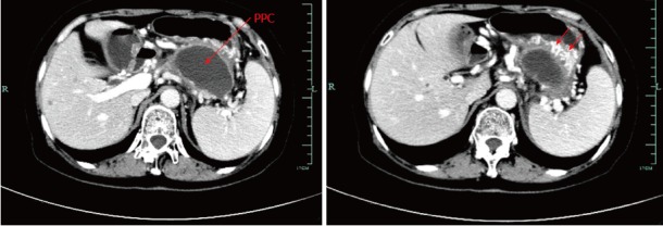
Figure 1.

Pre-procedural contrast-enhanced computed tomography image. Computed tomography (CT) scan of the upper abdomen images revealed a 7.4 cm × 6.2 cm pancreatic pseudocyst in the tail of the pancreas, which was in close contact with the posterior wall of the stomach. Notably, splenic vein occlusion, splenomegaly and gastric varices were also observed.
