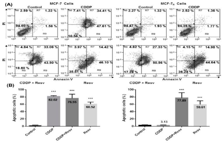
Figure 5.
Resv overcome CDDP-resistance and induces apoptosis in MCF-7R cells. (A) MCF-7 and MCF-7R cells were treated with a DMSO–ethanol vehicle as control or CDDP (6 μM) with or without Resv (100 μM) for 48 h and were double-stained with Annexin V and propidium iodide (PI) followed by flow cytometry analysis to determine apoptotic cells. The viable cells are located in the lower left quadrant (double negative with Annexin V–/PI–). Apoptotic cells (Annexin V+/PI–) appear in the lower right (early apoptosis) and upper right (late apoptosis) quadrant of data plots. Data are presented as percentage of the cell population. (B) The combined results of three independent cytometry analyses depicting the mean levels of total apoptotic cells are shown. Results are presented as the means ± SD. *** p < 0.001 by one-way ANOVA followed by Dunnett’s Multiple Comparison test.

