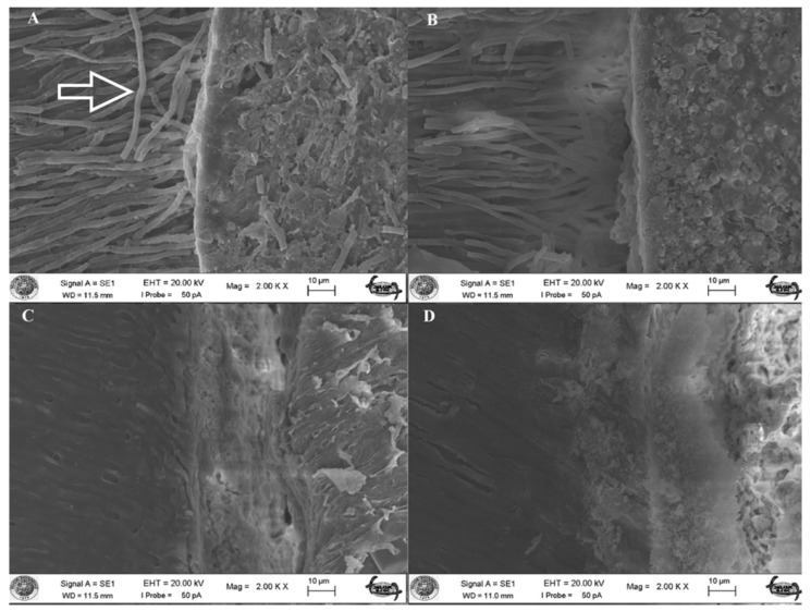
Figure 1.
(A) Scanning electron micrograph (×2000) of a sample in group 1 (AH/CLC). Radicular dentinal wall is densely covered with AH Plus sealer and resin tags (indicated with an arrow) occluding the dentinal tubules; (B) Scanning electron micrograph (×2000) of a sample in group 3 (AH/C). Dentinal wall is partly covered with the sealer and tags protruding from the sealer; (C) Scanning electron micrograph (×2000) of a sample in group 2 (ES/CLC). The area is covered with the sealer but there is no visible sealer tags in dentinal tubules; (D) Scanning electron micrograph (×2000) of a sample in group 4 (ES/C) where there is almost debonding of the sealer from the radicular dentinal wall and no sign of tag elements.

