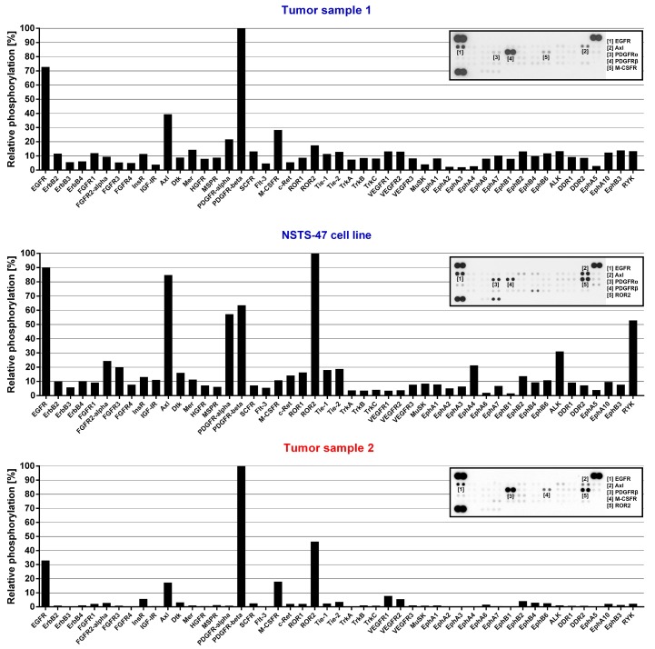
Figure 1.
Phospho-receptor tyrosine kinases (RTK) array analysis. The relative phosphorylation of 49 RTKs was analyzed in tumor tissue obtained from the boy when he was 3.5 months old (Tumor sample 1), in the NSTS-47 cell line (derived from a tumor tissue of the boy obtained when he was 1 year and 7 months old) and in the tumor tissue of his 8-year-old sister (Tumor sample 2). platelet-derived growth factor receptor beta (PDGFR-beta) and epidermal growth factor receptor (EGFR) exhibited high levels of phosphorylation in all cases. Phosphorylation in NSTS-47 cells was measured after 24 h of serum-free cultivation. The array images captured using X-ray film are shown for each sample, and the five most phosphorylated receptor tyrosine kinases (RTKs) are marked. The upper part of the figure (Tumor sample 1) was already published in our previous case report [8] under the Creative Commons Attribution 4.0 International License.

