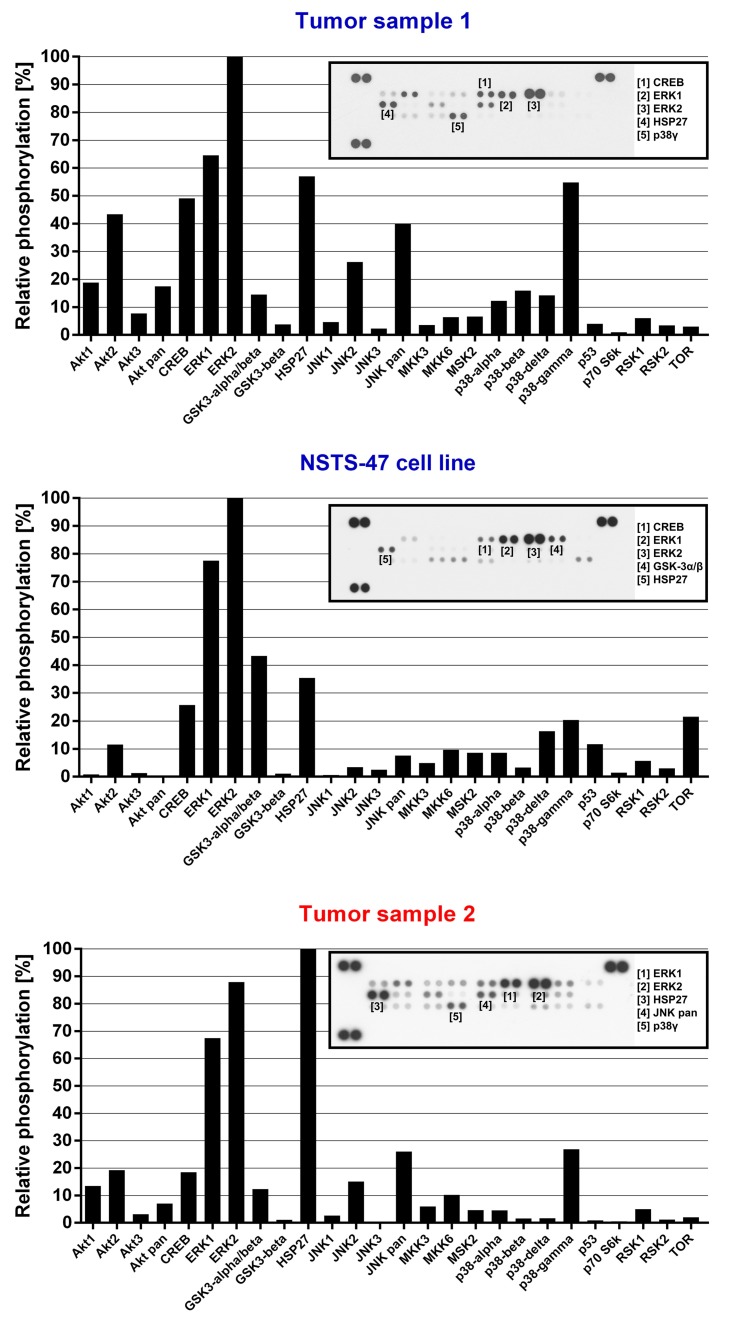
Figure 2.
Phospho-mitogen-activated protein kinase (MAPK) array analysis. The relative phosphorylation of 26 signaling proteins, including 9 MAPKs, was detected in tumor tissue obtained from the boy when he was 3.5 months old (Tumor sample 1), in the NSTS-47 cell line (derived from a tumor tissue of the boy obtained when he was 1 year and 7 months old) and in the tumor tissue of his 8-year-old sister (Tumor sample 2). ERK1/2 exhibited high levels of phosphorylation in all cases. Phosphorylation levels in NSTS-47 cells was measured after 24 h of serum-free cultivation. The array images captured using X-ray film are shown for each sample, and the five most phosphorylated proteins are marked.

