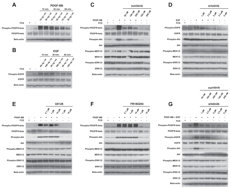
Figure 4.
Analysis of protein phosphorylation. (A) PDGFR-beta phosphorylation is increased in response to PDGF-BB. Cells were stimulated for 15, 30 or 60 min using two different concentrations (10 ng/mL and 30 ng/mL) of PDGF-BB. (B) EGFR phosphorylation is increased in response to epidermal growth factor (EGF). Cells were stimulated for 15, 30 or 60 min using two different concentrations (40 ng/mL and 100 ng/mL) of EGF. (C) Sunitinib was able to decrease PDGFR-beta and Akt phosphorylation but not MEK1/2 and ERK1/2 phosphorylation. (D) Erlotinib decreased EGFR and Akt phosphorylation but had no effect on MEK1/2 and ERK1/2 phosphorylation. (E) U0126 treatment did not decrease MEK1/2 phosphorylation. (F) FR180204 treatment did not cause any changes in ERK1/2 phosphorylation. (G) The combination of sunitinib and erlotinib decreased PDGFR-beta, EGFR and Akt phosphorylation, but MEK1/2 and ERK1/2 phosphorylation was not affected.

