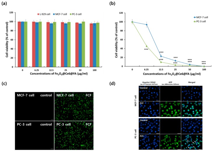
Figure 4.
Biocompatibility and photodynamic anticancer activities of chlorin e6 and folic acid-conjugated magnetite (Fe3O4-Ce6-FA) nanoparticles. (a) Cytotoxicity and (b) phototoxicity of Fe3O4-Ce6-FA nanoparticles in MCF-7 (breast adenocarcinoma) and PC-3 (prostate adenocarcinoma) cell lines. Quantitative data are expressed as the mean ± standard deviation (n = 4), and the statistical comparisons were evaluated using Student’s t-test. Significant differences were indicated by p < 0.05 (*** p < 0.0005 vs. control). (c) Images of MCF-7 and PC-3 cells after staining with fluorescein isothiocyanate-conjugated Annexin V (Annexin V-FITC) thus demonstrating the membrane translocation of the cells. The green fluorescence signal was produced by Annexin V-FITC. “FCF” represents Fe3O4-Ce6-FA nanoparticles. Scale bar = 50 μm. (d) Nuclear fragmentation and caspase-3/7 activity in MCF-7 and PC-3 cells. The cells were stained with Hoechst 33342 to detect nuclear fragmentation and CellEvent Caspase-3/7 Green Detection reagent to detect caspase-3/7 activity after 6 h post photodynamic therapy at 20 mW for 30 min. Arrows represent apoptotic bodies of cells. Scale bar= 30 μm.

