Figure 1.
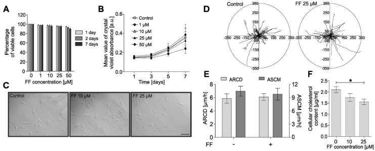
Fenofibrate does not affect the viability, proliferation, morphology, or motility of human bronchial fibroblasts (HBFs). (A) HBFs derived from asthma patients were cultured in the absence or presence of fenofibrate (FF; 1–50 µM) for one, two, and seven days, and the number of viable cells was detected using the fluorescein diacetate/ethidium bromide (FDA/EtBr) test. (B) Proliferation of HBFs after FF (0–50 µM) administration was measured after one, three, five, and seven days of cell cultivation, presented as a mean value of crystal violet absorbance. (C) Representative images of HBF morphologies after treatment with FF for 48 h (integrated modulation contrast, IMC). Scale bar = 25 µm. (D,E) HBFs were cultured in the absence or presence of FF (25 μM) for 48 h. The results of time-lapse monitoring of HBF movement are presented as circular diagrams, with the starting point of each trajectory situated at the plot center and column charts summarizing the effect of FF on the average speed of cell movement (ASCM; µm/h) and the average rate of cell displacement (ARCD; µm) parameters. (F) Intracellular cholesterol levels of HBF populations (n = 10) were measured with an Amplex Cholesterol Assay Kit after seven days of FF treatment (10 or 25 µM) in three independent experiments and are shown on a graph. Data are the mean ± standard error of the mean (SEM) of six independent experiments. Statistical significances were tested using one-way (A,C,F) or two-way ANOVA (B) with the Bonferroni multiple comparison post hoc test; * p ≤ 0.05.

