Figure 2.
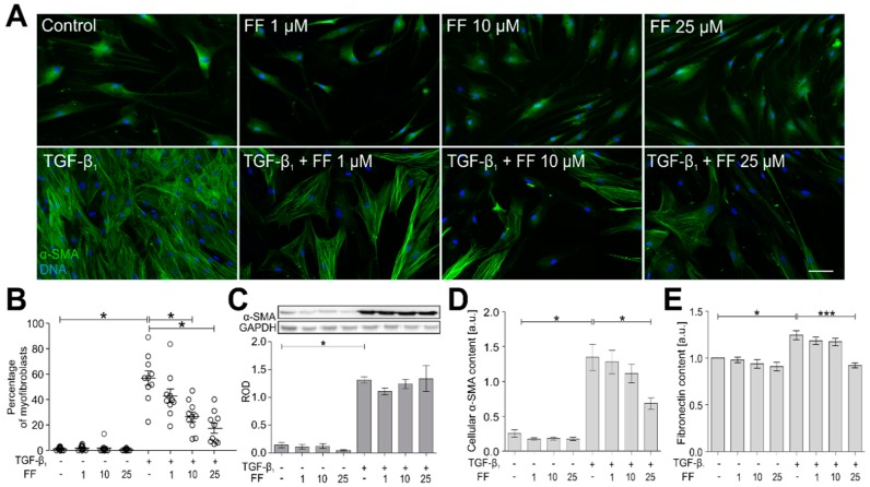
Fenofibrate attenuates the TGF-β1-induced phenotypic transition of HBFs into myofibroblasts. (A) HBFs were cultured in control conditions or in Dulbecco’s modified Eagle medium (DMEM) supplemented with TGF-β1 (5 ng/mL) in the absence or presence of FF (1–25 μM) for seven days. Then, the cells were fixed with 3.7% formaldehyde, permeabilized, and immunostained for α-smooth muscle actin (α-SMA; green) and DNA (blue) as shown on representative images. Scale bar = 25 µm. (B) The fraction of cells with prominent α-SMA+ stress fibers in HBF populations (n = 10) was determined using fluorescence microscopy in three independent experiments. (C) Analyses of α-SMA content were carried out in total cell lysates from HBFs cultured as in (A) using immunoblotting. Human glyceraldehyde 3-phosphate dehydrogenase (GAPDH) was used as a loading control. The effect of fenofibrate on the α-SMA levels in the TGF-β1-treated HBFs is presented as a bar graph and shows densitometric quantification of Western blots. Data are the mean ± SEM of five independent experiments in triplicate. (D,E) α-SMA and fibronectin contents, respectively, were defined using in-cell ELISA, and the results are presented as the mean value of absorbance (450 nm) reflecting the protein content. Data represent the mean ± SEM carried out on HBFs (n = 10), each in triplicate. Statistical significances were tested using one-way ANOVA with the Bonferroni multiple comparison post hoc test; * p ≤ 0.05, *** p ≤ 0.001. Note that fenofibrate significantly inhibits the formation of α-SMA+ stress fibers in the TGF-β1-treated HBFs in a dose-dependent manner, and concomitantly, has a slight impact on the total α-SMA level.

