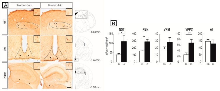
Figure 1.
Linoleic acid deposition on the tongue induces c-Fos expression in the major cerebral structures of the canonical gustatory pathway. (A) Typical photomicrographs of the NST, Arc, and Hipp, showing c-Fos immunoreactivity in mice subjected to oral stimulation with linoleic acid or xanthan gum (XG, 0.3%, w/v) to mimic the texture of lipids. The dotted lines circumscribe the regions of interest. Arrowheads point to representative c-Fos immunopositive nuclei. The boxes show higher magnification (×4) of representative c-Fos immunopositive nuclei. Scale bar, 100 µm. The square windows indicate the area shown in the photomicrographs. (B) Bar graph representation of the density of c-Fos immunopositive cells (number of c-Fos positive cells/mm2) in mice subjected to oral stimulation with linoleic acid (LA) or xanthan gum (XG). Values are means ± standard error of the mean (SEM); n = 6; for each structure studied, treatment effects on c-Fos expression were assessed using unpaired one-sided t-tests. *, p < 0.05; **, p < 0.01. AI, agranular insular cortex; Arc, arcuate nucleus; Hipp, hippocampus; NST, nucleus of the solitary tract; PBN, parabrachial nucleus; VPM, ventral posteromedial thalamic nucleus; VPMPC, ventroposterior medialis parvocellularis.

