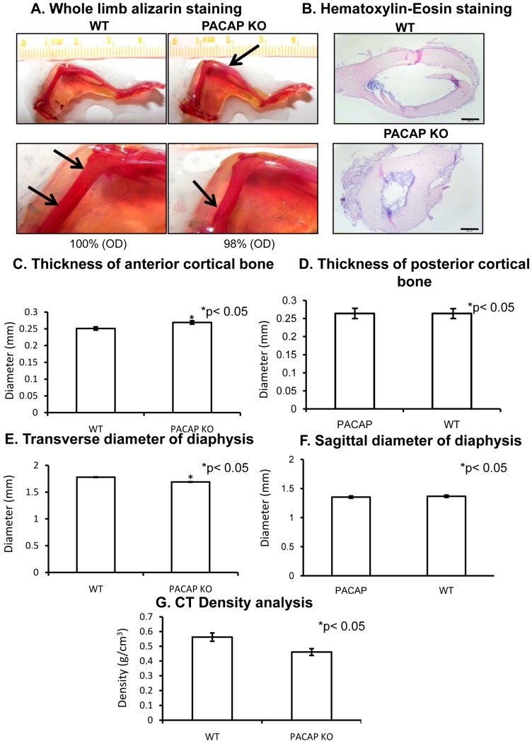
Figure 1.
Morphological analysis of hind limbs of wild type (WT) and pituitary adenylate cyclase activating polypeptide (PACAP) knockout (KO) mice. Whole limb alizarin red staining (A), optical density (OD) was determined in 1 cm distal part of the femur. Hematoxilin-eosin (HE) staining (B) to visualize the histological differences. Original magnification was 4×. Scale bar, 500 µm. CT analysis (C–G) of mouse femurs. Representative data of 5 independent experiments. Asterisks indicate significant (* p < 0.05) difference in thickness of cortical bone or in the diameter of diaphysis compared to the respective control.

