Figure 1.
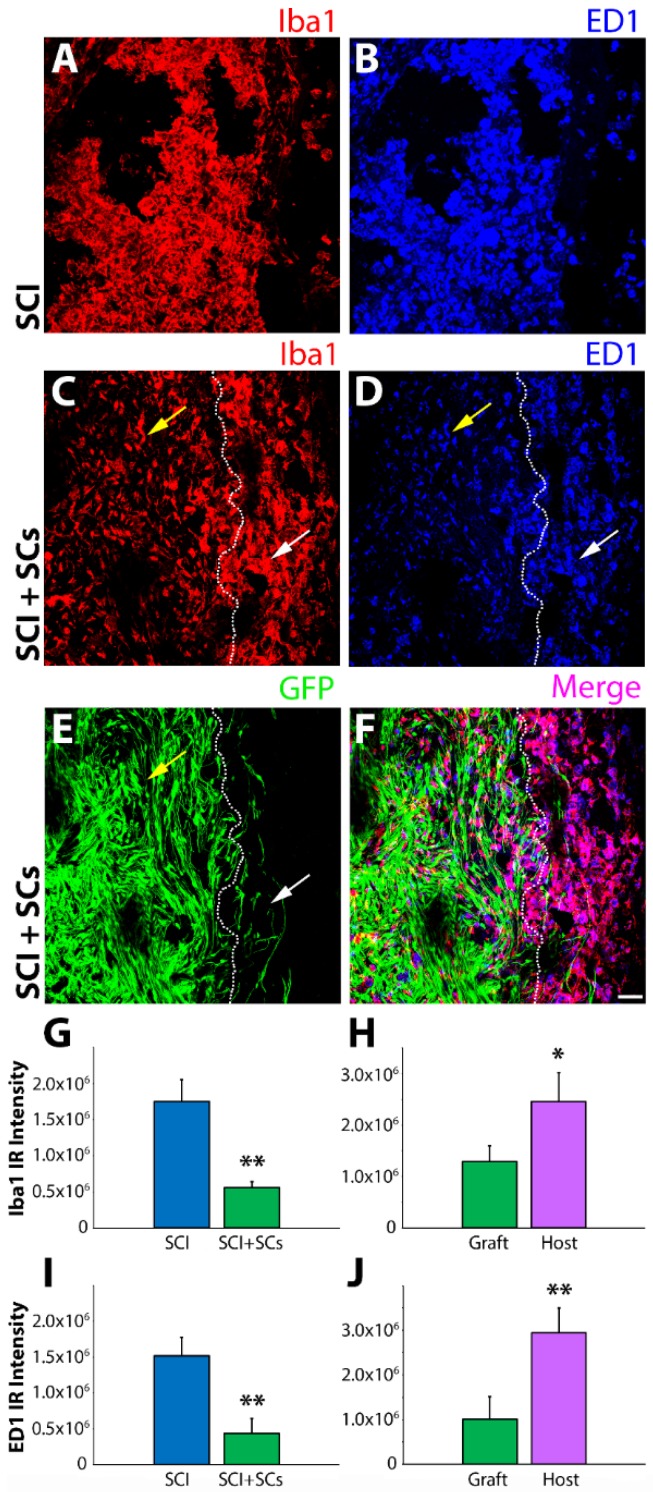
Immunoreactivity for ionized calcium-binding adapter molecule 1 (Iba1) and cluster of differentiation molecule 68 (CD68) was reduced within the lesion–Schwann cell (SC) implant of transplanted animals after spinal cord injury (SCI). Confocal micrographs of horizontal spinal cord sections from SCI control and SCI, enhanced green fluorescent protein (EGFP-SC)-transplanted animals (n = 4) at 2 weeks post-injury (1 week post-transplantation) immunostained for Iba1 (red) and CD68 (blue). In SCI control tissue, there was significant infiltration of both Iba1 and CD68 immune cells within the lesion (A,B). In contrast, in EGFP-SC-transplanted animals, the numbers of Iba1 and CD68 immune cells was greatly attenuated within the lesion–SC implant (C–F). Quantification of fluorescent intensity showed that EGFP-SC transplantation led to reductions in both Iba1 (G) and CD68 (I) that were more pronounced within the lesion than in adjacent host tissue (H,J). Results expressed as mean ± standard error of the mean (SEM). Statistical significance indicated at * p < 0.05 and ** p < 0.01 compared with SCI controls. Images were acquired at 20× objective magnification. Yellow arrows indicate the lesion-SC implant and white arrows the perilesional area. Scale bar = 50 μm.

