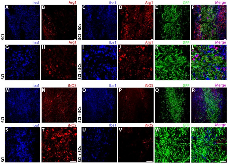
Figure 7.
Lesional and perilesional changes in Arg1 and iNOS were observed after SCI and SC transplantation. Immunohistochemical staining showed a pronounced increase in Arg1 immunoreactivity (red) from SCI controls (A,B,G,H) to EGFP-SC-transplanted (green) animals (C,D,I,J). The increase in Arg1 was most pronounced in the perilesional region and adjacent host spinal cord tissues (E,F,K,L). In contrast, compared with SCI controls (M,N,S,T), iNOS immunoreactivity (red) was dramatically reduced after EGFP-SC transplantation (O,P,U,V), being virtually absent from the lesion–SC implant (Q,R,W,X). Total innate immune cells identified using Iba1 (blue). Triple staining of EGFP, Iba1 and either Arg1 or iNOS are shown in the merged images (pink; (F,L,R,X)). Images were acquired at 10× (A–F, M–R) and 40× (G–L, S–X) objective magnifications. Scale bar = 50 μm.

