Figure 2.
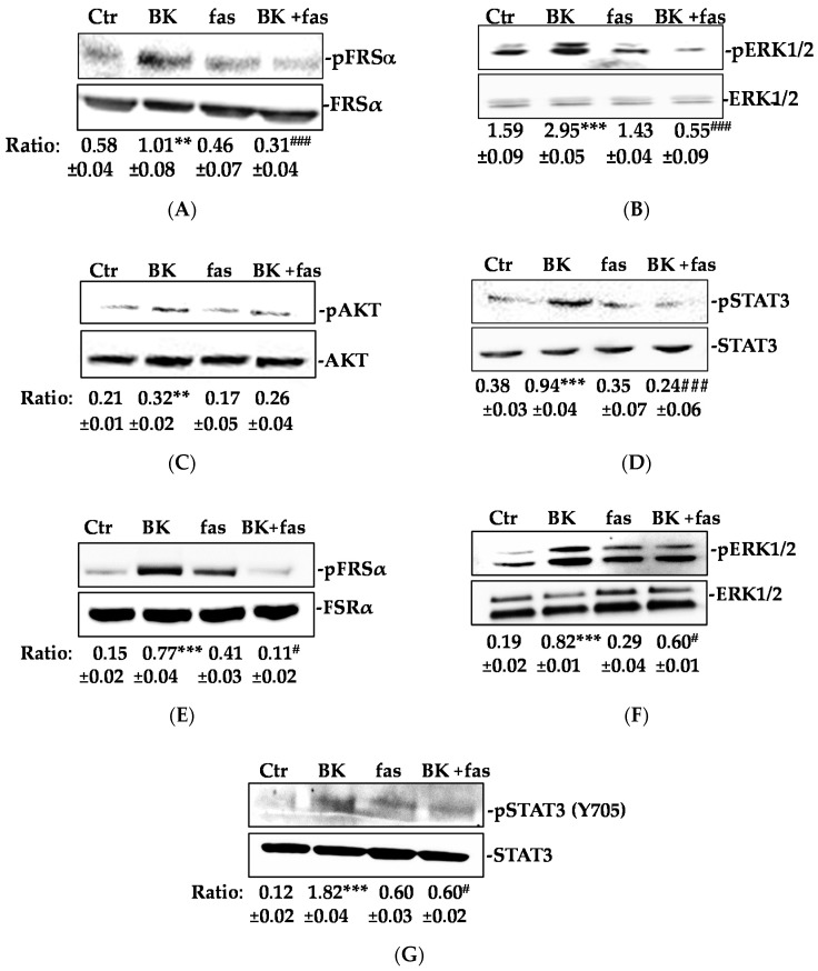
BK/B2R activates FGFR-1 signaling. (A) FRSα, (B) ERK1/2, (C) AKT, and (D) STAT3 phosphorylation were evaluated using western blot analysis in HUVEC treated with fasitibant (fas, 1 μM, 30 min), then stimulated with BK (1 μM) for 15 min. (E) FRSα, (F) ERK1/2, and (G) STAT3 phosphorylation were evaluated using western blot analysis in HREC treated with fasitibant (fas, 1 μM, 30 min), then stimulated with BK (1 μM) for 15 min. Results were normalized to FRSα, ERK1/2, AKT, and STAT3, respectively. The results presented are representative of three independent experiments (n = 3) with similar results. Quantification was expressed as an arbitrary density unit (ADU). ** p < 0.01; *** p < 0.001 vs. Ctr; # p < 0.05; ### p < 0.001 vs. BK treated cells.

