Figure 2.
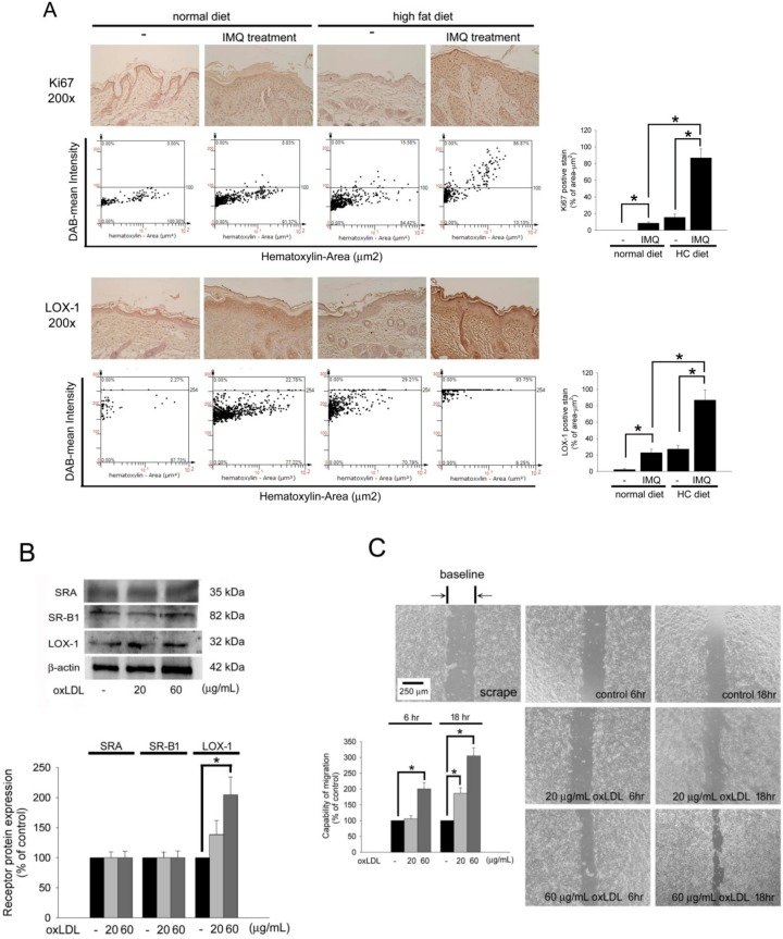
OxLDL increases keratinocytes migration and LOX-1 expression. (A) The skin slides were stained with anti-Ki67 or anti-LOX-1 antibodies. The images were acquired at a 200× magnification and quantified using TissueGnostics TissueFAXS & HistoFAXS System (TissueGnostics, Vienna, Austria). (B) Hacat cells were treated with 20 or 60 µg/mL of oxLDL, and the total protein was analyzed to assay the SR-A, SR-B1, and LOX-1 expression. β-actin was used as a loading control. The density of each band was quantified by a densitometer. (C) Wound healing assays for evaluating the effect of oxLDL on Hacat cell migration. Hacat cells migrating to the denuded area were counted based on the black baseline. Hacat cells were cultured with oxLDL for 24 h before wound scraping using a pipette tip. The photographs were taken 6 and 18 h after wound scraping (×40). The Hacat cells that migrated into the denuded area (double arrows indicate the denuded area) were analyzed. The magnitude of Hacat migration was evaluated by counting the migrated cells in six random clones under a high-power microscope field (×100). The results were expressed as the mean SD; * p < 0.05 was considered statistically significant.

