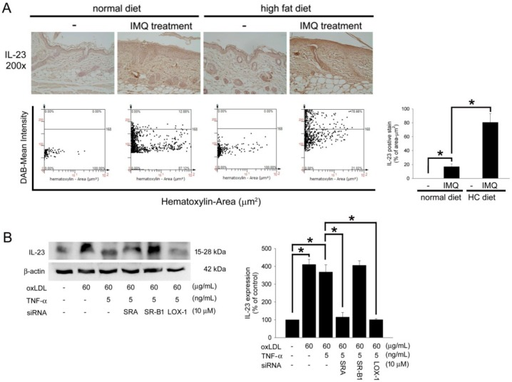
Figure 4.
HC-diet aggravated the IL-23 expression in IMQ-treated B6.129S2-Apoetm1Unc/J mice and oxLDL induced IL-23 expression mediating by LOX-1 in TNF-α-stimulated Hacat cells. (A) The representative photos of the mice received IMQ treatment in a normal chow diet or HC-diet groups. The skin slides were stained with anti-IL-23 antibody. The images were acquired at a 200× magnification and quantified using TissueGnostics TissueFAXS & HistoFAXS System (TissueGnostics, Vienna, Austria). The density of IL-23 expression was presented as a bar graph (right). (B) Hacat cells were incubated with oxLDL or oxLDL plus TNF-α for 24 h with or without preincubation of specific competitive scavenger antibodies. Total proteins were extracted and western blot analysis was performed. β-actin was used as a loading control. The density of each band was quantified by a densitometer. All results were expressed as the mean ± SD. A * p < 0.05 was considered statistically significant.

