Figure 1.
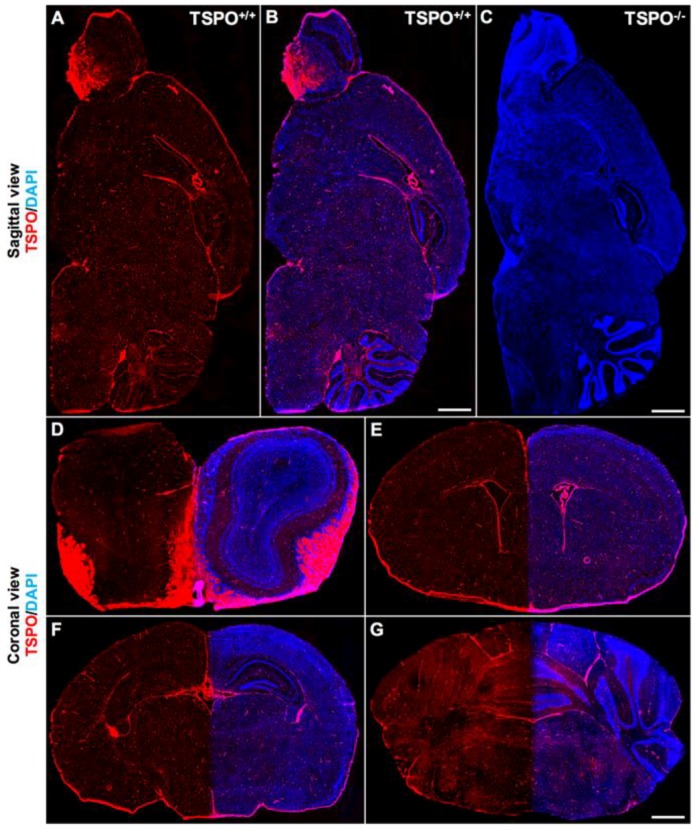
Global immunofluorescence overview of TSPO (translocator protein 18 kDa) expression across major brain regions. (A) Sagittal section from a wildtype mouse (TSPO+/+) demonstrates widespread TSPO expression (red) across all brain regions using a specific antibody against TSPO (anti-PBR, ab109497). (B) Sagittal section from a wildtype mouse with TSPO expression in red and 4’,6-diamidino-2-phenylindole (DAPI) in blue highlighting structural features. Strong TSPO immunoreactivity is present in the olfactory bulb, the choroid plexus and the ependyma of the ventricular system, and cerebellum. (C) Sagittal section from a TSPO knockout mouse (TSPO−/−) confirming the absence of TSPO expression (DAPI in blue). (D) Coronal view of the olfactory bulb demonstrates strong TSPO expression in the olfactory nerve layers and glomeruli, and concentrated expression in the subependymal zone. (E) TSPO expression is present in the subventricular zone and the ependymal cells of the ventricles and choroid plexus. (F) Coronal view of the hippocampal region with discernible TSPO expression observed in the dentate gyrus. (G) Cerebellar/brainstem TSPO expression is strongly present in the molecular layer of the cerebellar cortex and fiber tracts of the brainstem. Scale bars = 500 µm.

