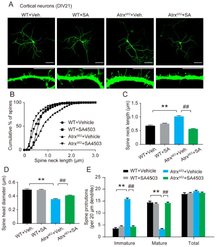
Figure 2.
Treatment with SA4503 ameliorates dendritic spine abnormality in cultured AtrxΔE2 neuron. (A) Representative images of EGFP-transfected cortical neurons at DIV21. Neurons are stained for anti-GFP. Images in the bottom panels are enlarged from each dendrite. Scale bars: top panels, 100 µm; bottom panels, 10 µm. (B) Relationship between cumulative percentage of spines and spine length. p < 0.01 in WT versus AtrxΔE2 neurons and in AtrxΔE2 versus SA4503-treated AtrxΔE2 neurons by the Kolmogorov–Smirnov test. (C) Data show the number of spines. ** p < 0.01 versus vehicle-treated WT neurons; ## p < 0.01, versus vehicle-treated AtrxΔE2 neurons by one-way ANOVA with post hoc Tukey’s test; F (3, 1494) = 89.38. (D) Data show the spine-head diameter. ** p < 0.01 versus vehicle-treated WT neurons; ## p < 0.01, versus vehicle-treated AtrxΔE2 neurons by one-way ANOVA with post hoc Tukey’s test; F (3, 76) = 76.38. (E) Spine protrusions per 20 µm dendritic length. ** p < 0.01 versus vehicle-treated WT neurons; ## p < 0.01, versus vehicle-treated AtrxΔE2 neurons by two-way ANOVA with post hoc Bonferroni’s test; F (3, 228) = 2.549, p = 0.0566 (group); F (2, 228) = 1008, p < 0.01 (spine protrusions), F (6, 228) = 243.6, p < 0.01 (interaction between group and spine protrusions); n = 20 neurons for each group. The experiments were repeated three times with similar results. WT, n = 360 spines; SA4503-treated WT, n = 367 spines; AtrxΔE2, n = 384 spines; SA4503-treated AtrxΔE2, n = 370 spines. Each bar represents the mean ± SEM. Abbreviations: Veh., vehicle; SA, SA4503.

