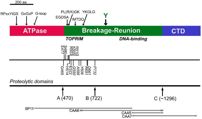
Figure 1.
Schematic representation of the domain arrangement of human topoisomerase IIβ. The ATPase domain is shown in red, the breakage-reunion domain in green and the C-terminal domain in blue. The positions of conserved motifs are marked above. The line underneath shows the location of site-directed mutations reported by West et al. [7] (above) and mutations selected for drug resistance reported in Leontiou et al. [8] (below). The locations of inter-domain hinge regions elucidated by limited proteolysis reported in Austin et al. [9] are also shown. At the bottom of Figure 1, the locations of the initial partial cDNA clones encoding portions of TOP2B are shown [5]; SP11, and CAA5, clone CAA6, confirming that CAA5 and SP11 were part of the same gene, are also shown [10,11].

