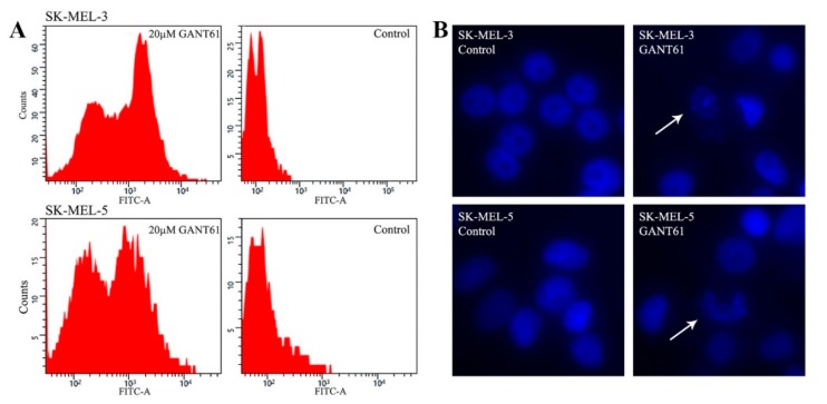
Figure 3.
(A) TUNEL assay detecting apoptosis in two cell lines. Cells were seeded on 60-mm dishes, and the next day, 20 μM GANT61 was added. The normal medium was replaced in controls. After three days, the majority of cells treated with GANT61 detached in both SK-MEL-3 and SK-MEL-5 cells. Both detached and remaining attached cells were used for analysis. FITC fluorescence clearly shows massive apoptosis in GANT61-treated cells. The percentage of the apoptotic and nonapoptotic cells were calculated using ImageJ software (National Institutes of Health, Bethesda, MD, USA). The results of cell quantification were as follows. SK-MEL-3 cells treated with GANT61: apoptotic cells 62.62%, nonapoptotic cells 37.38%; SK-MEL-3 controls: apoptotic cells 0.4%, nonapoptotic cells 99.6%. SK-MEL-5 cells treated with GANT61: apoptotic cells 51.97%, nonapoptotic cells 48.03%; SK-MEL-5 controls: apoptotic cells 4.18%, nonapoptotic cells 95.82%. No cell cycle blockade was observed. (B) Fluorescence showing apoptotic nuclei in the same cells as in (A), treated equally with GANT61 or untreated (control cells). Cells were mounted in a medium containing DAPI and documented by fluorescence. Magnification: 200×. Arrows show apoptotic nuclei.

