Figure 5.
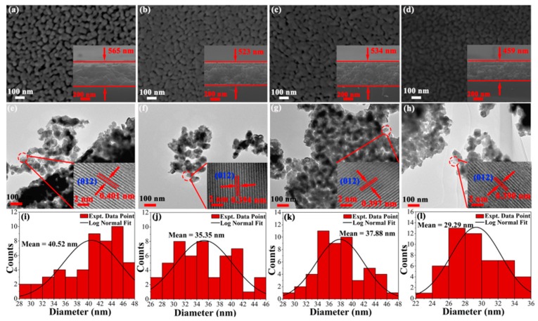
SEM images of surface morphologies for (a) BFO (b) BHFO (c) BFMO (d) BHFMO thin film and the insets in (a–d) show cross-sectionals of all thin films; (e–h) low magnification TEM images of BFO, BHFO, BFMO, BHFMO thin films and the insets in (e–h) show high resolution TEM images of all thin films; Histograms regarding particle size distributions of (i) BFO (j) BHFO (k) BFMO (l) BHFMO thin film, respectively.

