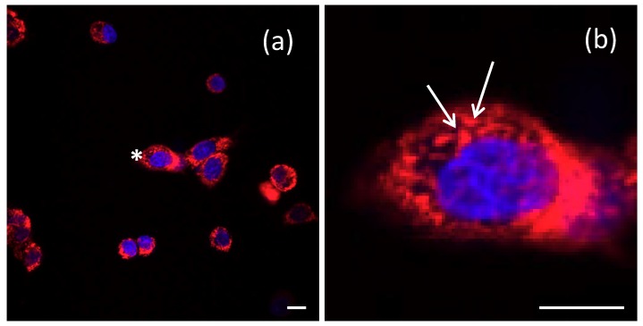
Figure 5.
Intracellular localization of dextran 70,000-coated SPIO-NPs labelled with rhodamine 123, in C6 cells after 24 h of incubation, is shown by confocal microscopic images (a). The image on the right (b) zooms into the cell labeled with an asterisk in (a) to illustrate the dotted labelling (arrows), which presumably indicates endosomal–lysosomal uptake. Nuclei stained with Hoechst 33342 in blue. Scale bars 20 µm.

