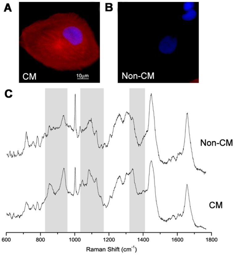
Figure 6.
Typical immunostaining images of: (A) individual cardiomyocytes (CM); and (B) non-cardiomyocytes (non-CM) derived fromhuman embryonic stem cells (hESCs). Cell nuclei DAPI (blue); cardiac a-actinin (red). (C) Raman spectra of the same cells. Reprinted with permission from [67]. Copyright 2018, Elsevier.

