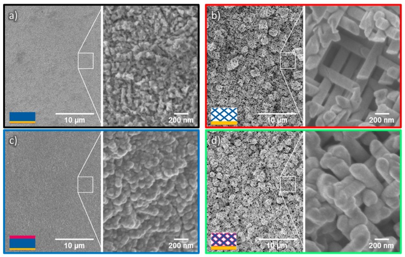
Figure 6.
Low (left) and high (right) magnification SEM images, displaying (a) a ZnO film exhibiting a rough surface and spike-like features; (b) a ZnO nanowire network; (c), a ZnO film coated with 20 nm TiO2 and (d) a ZnO/TiO2 core-shell nanowire network. In the insets’ schemes, the colors present the different materials: gold (yellow), ZnO (blue), and TiO2 (pink).

