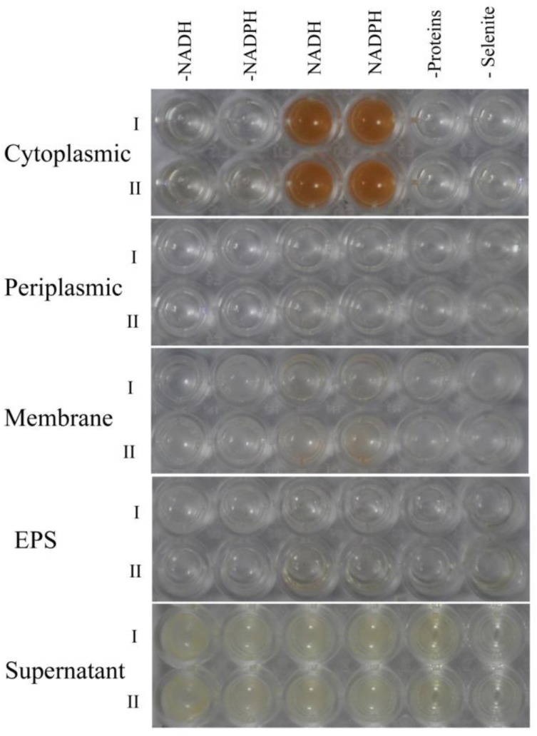
Figure 8.
Selenite reduction assay on different subcellular fractions (cytoplasmic, periplasmic, and membrane), supernatant, and exopolysaccharide. All experiments were performed in duplicate (indicated by roman numbers), with addition of 5.0 mM SeO32− and 2.0 mM NADH or NADPH. While 3 following control negatives were performed: without protein fractions or supernatant or EPS, without selenite, without NADH or NADPH.

