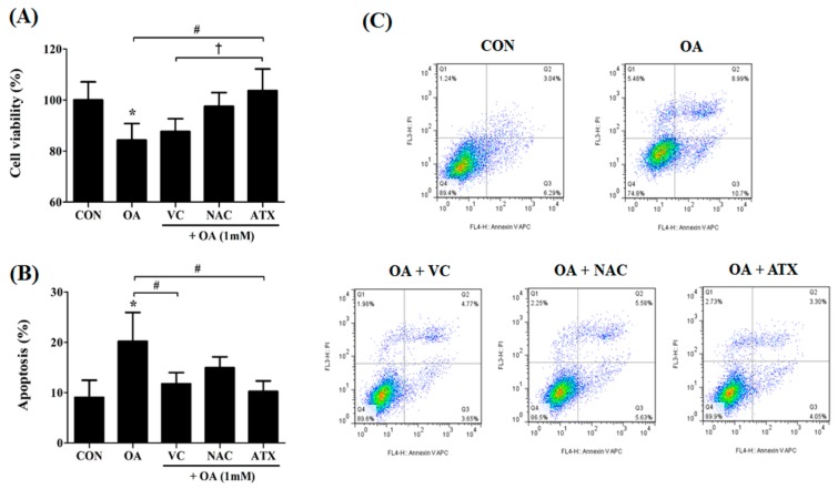
Figure 6.
Effects of the antioxidants on cell apoptosis resulting from OA-induced steatosis. The cell viability (%) after antioxidant treatments in the OA-treated cells was investigated using the CCK-8 kit (A), and apoptotic cells (%) were counted by the sum of the percentages of annexin V-stained cells and dual stained cells of Annexin V and PI using FACS analysis (B). Gating images for the apoptotic cells are shown in (C). Data are represented as mean ± SD values (n = 4). Asterisk (*) indicates a significant difference compared with the control (p < 0.05). Sharp (#) indicates a significant difference among the experimental groups (p < 0.05). Dagger (†) indicates a significant difference compared with the each other among the antioxidants, VC, NAC, and ATX, using one-way ANOVA (p < 0.05).

