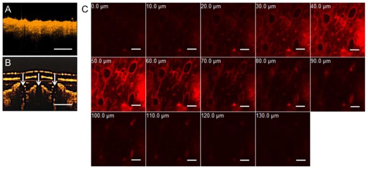
Figure 3.
In vitro transdermal delivery of R6G in mice via patches of gelatin/CMC MNs. (A) OCT image of mice skin before MNs insertion. (B) OCT image of mice skin after insertion. White arrows indicate the location of micro-disruption in the dermis. Scale bar = 500 μm. (C) The R6G-loaded gelatin/CMC MNs penetrate mice skin at certain depths after insertion for 60 min. Scale bar = 200 μm.

