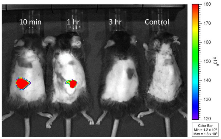
Figure 4.
In vivo fluorescence images of db/db mice at 10 min, 1 h and 3 h after treatment of FITC-insulin-loaded gelatin/CMC MN patches and unloaded (control) MN patches. The insertion sites of mice dorsal skin presented a strong fluorescent signal at 10 min indicating that the dissolving MNs rapidly released encapsulated FITC-insulin. The fluorescence intensity decreased at 1 h and disappeared at 3 h after application of MN patches.

