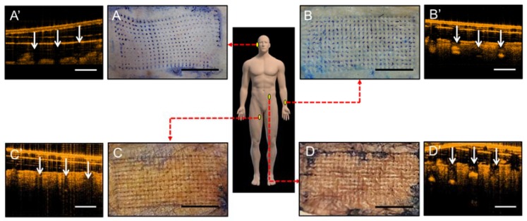
Figure 6.
In vitro insertion capability of patches of gelatin/CMC MNs applied to discarded human skins from various locations which were then stained with blue tissue-marking dye after patches removal. (A) Dorsal ear skin. (B) Ventral forearm skin. (C) Medial thigh skin. (D) Abdominal wall skin. Scale bar = 5 mm. (A’–D’) Respective OCT images of human cadaveric skin after MNs insertion. White arrows indicate the location of micro-disruption in the dermis. Scale bar = 500 μm.

