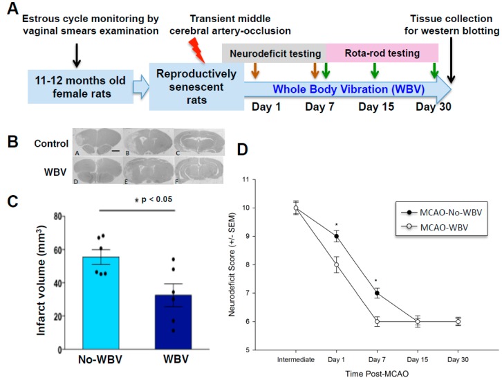
Figure 1.
(A) Experimental design. (B) Representative histological images of the brain (Bregma levels 1.2, −3.8, −5, 10X). (C) Geometric mean infarct volumes are compared between whole body vibration WBV and no-WBV groups. Post-ischemic WBV treatment shows reduced infarct volume as compared to the no-WBV group (* p < 0.05 as compared to no-WBV using student t-test). (D) Neurological deficit (ND) assessment scores were significantly improved in the WBV treated group as compared to no-WBV (* p < 0.05 as compared to no-WBV using Student Newman-Keuls).

