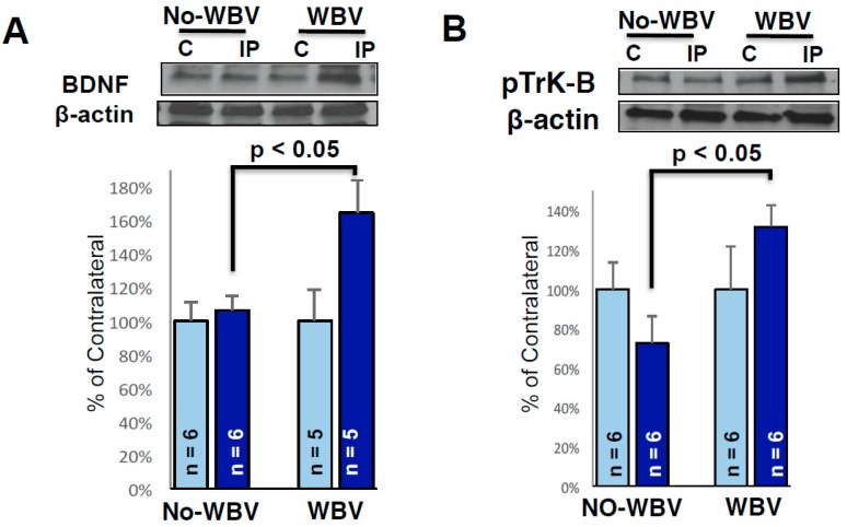
Figure 4.
Representative immunoblots showing the protein levels of BDNF and phosphorylated Trk-B in the peri-infarct area. β-actin (cytoskeletal), was used as a loading control. Densitometric analysis of scanned Western blots and expressed as percent of contralateral, showed baseline expression of BDNF (A) and phosphorylated Trk-B (B) proteins. Note the WBV treatment significantly increased BDNF and phosphorylated Trk-B in the peri-infarct area as compared to no-WBV (* p < 0.05 as compared to no-WBV using student t-test).

